About our Work

We are using molecular genetic approaches to study the development of neuronal cell types, with a particular focus on neuronal arbor formation in retinal neurons. Using Cre-loxP recombination, we specifically label individual neurons or neuronal cell classes, and visualize their morphology and connectivity. By combining conditional gene ablation with reporter gene replacement, we are able to visualize neurons in which specific genes have been ablated, and study the consequences of loss of gene function on the development of the neurons. Targeting specific neuronal populations through mouse genetic manipulations enables us also to study the roles of these neurons in visual circuitry.

Current research directions in the lab:

  • Developing methodologies for sparse genetic labeling of neuronal arbors, and gene function analysis.
  • Defining the transcriptional programs and molecular mechanisms of development and function of Retinal Ganglion Cell (RGC) types.
  • Tools for physiological and behavioral analysis of normal and pathological vision.
  • Using animal models to study the pathological mechanisms and therapeutic opportunities for blinding diseases.
  • Exploring repair and regeneration approaches in the visual system.
\

Molecular genetic
manipulations

Neuronal
Transcriptomes

Visual
Physiology/Behavior
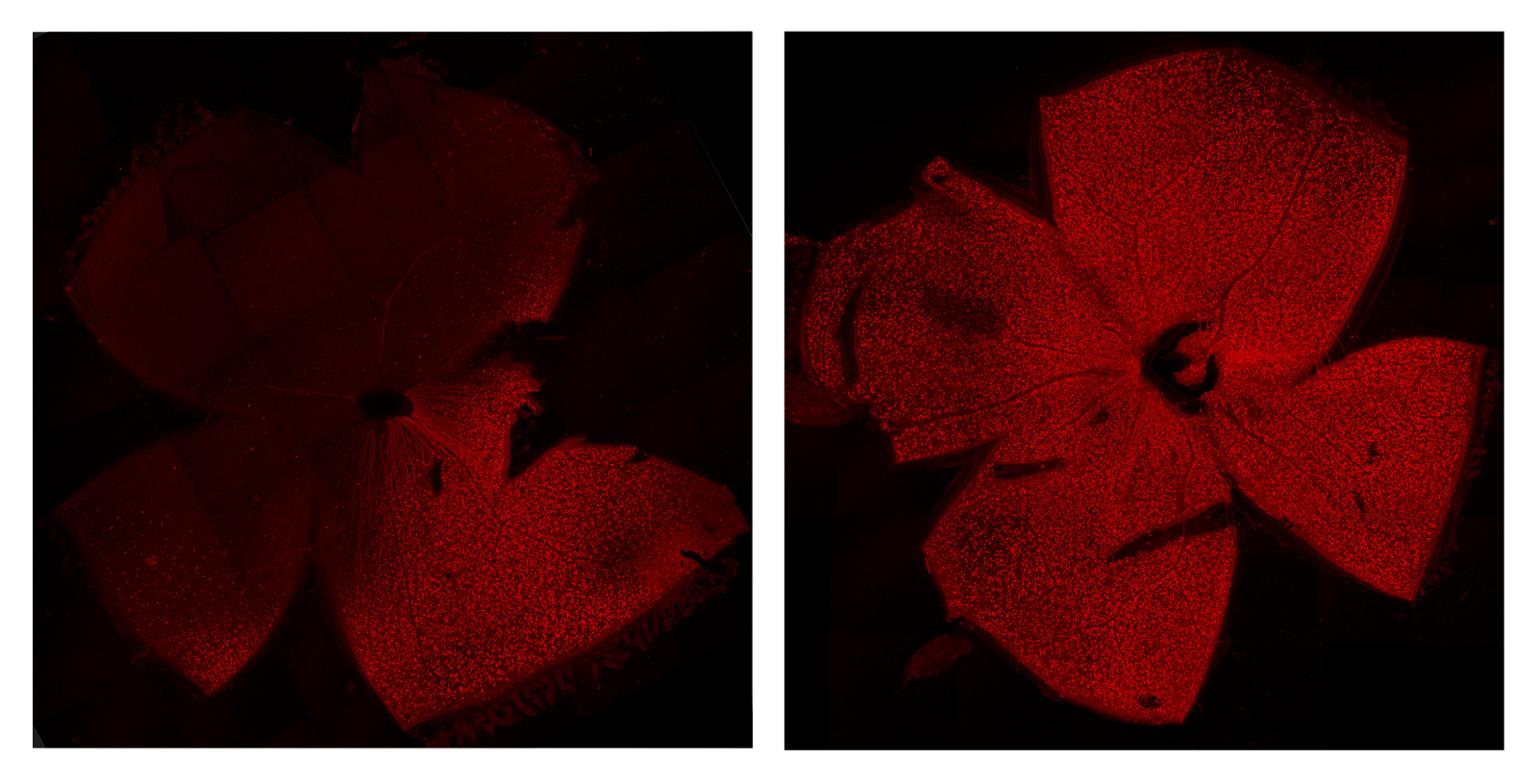
Left (ipsilateral) and right (contralateral) retinas, labelled using retrograde tracking of ChTB-A555 by injection of the left Superior Colliculus (Edwin Humberto Hodelin Maynard)
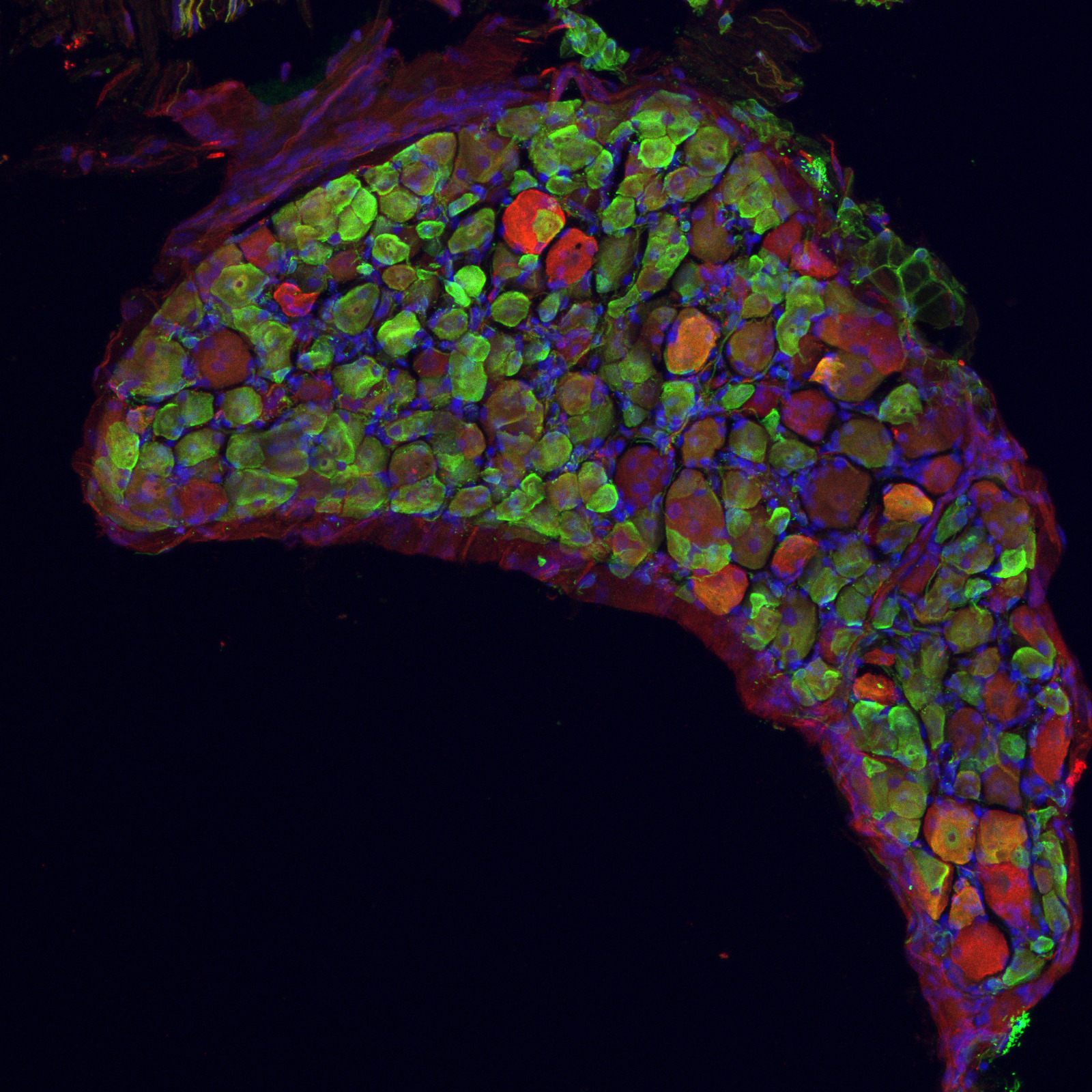
Dorsal Root Ganglion from a Tusc5eGFP/eGFP mouse labelled anti-eGFP (green) and anti-TrkC (red) (Dan Domocos, Anamaria Lazar, Ana-Maria Taranciuc)

Meet our Team

Current Lab members

Tudor C. Badea, MD, MA, PhD
Group Leader
Vladimir V. Muzyka, PhD
Postdoctoral Fellow
Anamaria Lazar, PhD
Master Student
Raluca Pascalau, MD
PhD Student
Edwin Humberto, MD
PhD Student
Diana Petre, MSc
Research Engineer
Ileana Daria Madan
Undergraduate student
Gisselle Fernndez Peña MD
Graduate student
Adina Teodora Rosca, Msc
Graduate student
Aloyma Veliz, MD
Graduate student

Former Lab members

Ana Maria Taranciuc (Sisman), MD
Clinical Laboratory resident
Vlad V. Vochitu, MD
Graduated Med. School 2023
Malina Barbu
Medical Student (6th year)
Oana Monica Leah
Medical Student (6th year)

Publications

View all Publications

Facilities Equipment

See more about Facilities Equipment

Current Funding and Projects

Established by Dr. Badea between NEI and Transilvania University for the purpose of supporting Visual Neuroscience and Ophthalmology research at UnitBv. NEI provided Dr. Badea's laboratory equipment (Microscopy, physiology, visual behavior), collection of genetically modified mouse lines (21 lines, about half generated by Dr. Badea at the NEI or Hopkins) and specific reagent collections (plasmids, probes, antibodies, cell lines). UnitBv financed the building and equipment of the BEAM facility, as well as most of the equipment of the Molecular Genetics and Neuroscience Laboratory. Student exchanges and common projects are planned.

We aim to understand the development, function, and pathology of central high acuity vision. In humans, high visual acuity is provided by the fovea, a specialized retinal structure, in which visual information flows in one-to-one fashion from cone photoreceptors to bipolar cells and high-resolution Retinal Ganglion Cells (RGCs). The modest progress in understanding the fovea and treating its disorders is due in part to the lack of a genetically accessible mammalian model. We have recently discovered an area of high visual acuity (Area Centralis, ArCe) in the mouse, and will explore its function and development, to establish it as a pathogenetic and therapeutic model for central vision and high visual acuity RGC defects. We will use unique genetically modified mouse lines we developed to identify the RGCs of the ArCe, determine their brain projections and participation in binocular vision, and study the developmental history and molecular mechanisms of ArCe formation. We will apply visually guided behavior tests to mice with genetic ablation of the ArCe in order to identify its involvement in visual function. The results will provide us with an animal model for studying pathogenetic mechanisms and therapeutic approaches for high visual acuity central visual defects. In addition, the gained insights will pave the way for current efforts in retina repair based on stem cell or reprograming efforts, by discovering the necessary developmental steps and molecular requirements.

Collaborations

  • Anand Swaroop - NEI - Genomics and retina transcriptomics
  • Samer Hattar (NIMH)/ Phyllis Robinson (UMBC) - ipRGCs
  • Horea Rus, Violeta Rus, Alexandru Tatomir (UMAB) & Sonia Vlaicu (UMF Iuliu Hatieganu) - role of RGC-32/Rgcc in inflammatory diseases.
  • Ching-Hwa Sung (Cornell Weill medical School) - functional analysis of retinal disease models
  • Daniel Kerschensteiner (WUSTL) - RGC types development and function
  • Greg Schwartz (Northwestern U.) - RGC physiology in health and disease
  • Brian Brooks (NEI) - Animal models of Coloboma and Albinism
  • Chai-An Mao (UTHSC) - transcriptional control of RGC type development
  • Sef Lucr. Dr. Vlad Monescu - Bioinformatics Analysis
  • Conf. Dr. Sorin Grigorescu - Vision-inspired AI approaches.
Image
18 January 2022

Retinal pigment epithelium-specific CLIC4 mutant is a mouse model of dry age-related macular degeneration

Image
04 March 2022

Cercetători români implicaţi în găsirea unui tratament pentru o boală de vedere extrem de gravă

Image
23 February 2022

Contribuţie importantă a unui medic cercetător de la Universitatea Transilvania, într-un proiect ...